Introduction
The human heart: It’s the powerhouse of our bodies, a tireless muscle that works day and night, pumping blood to every corner of our being. Understanding how this amazing organ works – its structure (anatomy) and its function (physiology) – is key to appreciating the incredible complexity and efficiency of the human body. In this Blog, we’ll explore the heart’s basic design, how its different parts fit together, and the remarkable process by which it keeps us alive. We’ll look at the chambers, valves, and blood vessels that make up this vital organ, and how they work together to circulate blood, delivering oxygen and nutrients while removing waste products. Get ready to discover the fascinating world of the human heart!
Table of Contents
Anatomy of the Heart
Location of The Heart
The heart is like a hard-working pump in our bodies, making sure that blood flows smoothly to all parts of the body. It is located in the middle of our chests, slightly tilted towards the left side. If you put your hand on your chest, just a little to the left side, you can feel the rhythmic beating of your heart. It is protected by our ribcage, which acts like a sturdy shield, keeping it safe. Its location is important because it allows the heart to easily pump blood to the rest of the body, ensuring that all our organs and tissues receive the oxygen and nutrients they need to function properly.
Structural Feature
Heart is a hollow, muscular structure that is mainly divided in two halves:
- Right side of the heart receives the Deoxygenated blood from the body and pumps it to the lungs for the oxygenation.
- Left side of the heart receives the Oxygenated blood from the lungs and pumps to the rest of the body.
The receiving chambers are known as Atrium and pumping chamber Ventricle.
We will deep dive into the heart walls, its layers, veins and chambers and will go through each one by one.
Heart Walls and Layers
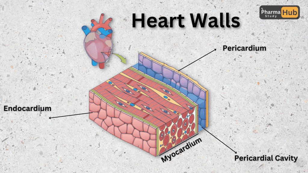
- Pericardium: It is the protective layer of the heart, it helps in the movement of the heart and helps in maintaining it in its position. it consist of 3 layers
- Fibrous Pericardium: It is a tough and elastic layer of the heart.
- Serous pericardium: It is the middle thin layer of the heart.
- Epicardium: It is the inner layer of the pericardium and it is connected to the heart.
- Myocardium: This is the layer which contracts to pump blood, it is a muscular and thick layer.
- Endocardium:This is the thin, smooth inner layer of the heart, made up of flattened epithelial cells, and it seamlessly continues as the endothelial lining of the blood vessels.
Chamber of the Heart
- Right Atrium: As we discussed above the atrium receives the blood, right atrium receive deoxygenated blood from 3 main veins:
- Superior vena cava: Brings deoxygenated blood from the upper body (head, arms, and chest).
- Inferior vena cava: Brings deoxygenated blood from the lower body (abdomen, pelvis, and legs).
- Coronary sinus: It Brings deoxygenated blood from the heart muscles.
- Right Ventricle: This Chamber receives blood from the right atrium and pumps the blood to the lungs for oxygenation of blood. This chamber is thicker than the atrium chamber, because this is the pumping chamber.
- Left Atrium: This chamber receives the oxygenated blood from the lungs and pumps it to the left ventricle.
- Left Ventricle: The chamber is full of oxygenated blood and it pumps to the whole body through the aorta, and then arteries deliver blood the whole body.
Heart Valves
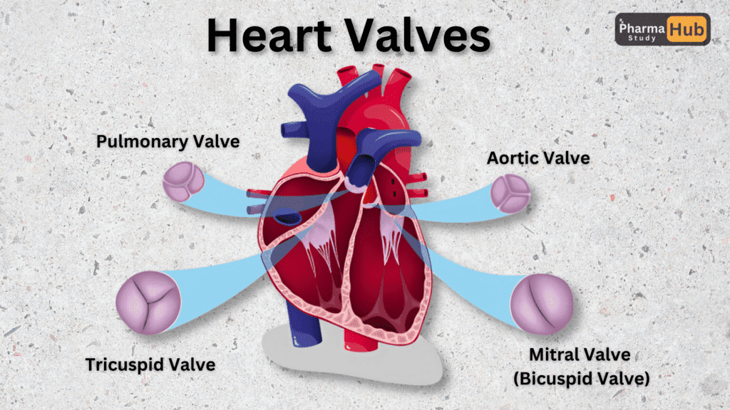
Valves are one-way passageways for blood flow, preventing blood flow from backing up when one heart chamber pumps blood to another chamber or to the lungs or body.
- Tricuspid Valves: These valves, also known as atrioventricular valves (AV valves), are located between the right atria and the ventricles, and are so named because they consist of 3 cups.
- Pulmonary Valves: This valve is found between our right ventricle and lungs, it prevents the return of blood pumped from the ventricle.
- Mitral Valves: This valve is also known as bicuspid valve, it is found between the left atrium and left ventricle. Its structure is like two cups, hence its name is bicuspid.
- Aortic Valves: This valve is found between our left ventricle and aorta. This valve prevents the backflow of blood that is sent to our body.
Major Blood Vessels
Arteries
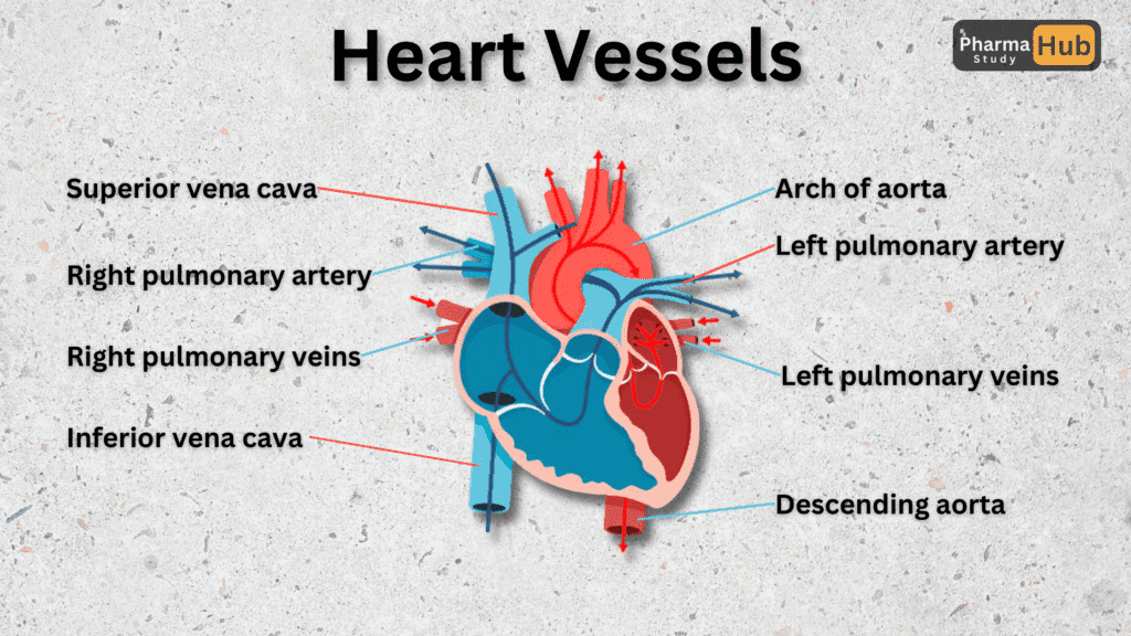
- Aorta: The largest artery in the body, it carries oxygen-rich blood from the left ventricle of the heart to the rest of the body.
- Coronary Artery: These arteries supply oxygen-rich blood directly to the heart muscle, ensuring it has the energy to pump effectively.
- Pulmonary Artery: The only artery that carries deoxygenated blood, it transports blood from the right ventricle to the lungs for oxygenation.
Veins
- Superior Vena Cava: A large vein that brings deoxygenated blood from the upper body (head, neck, arms, and chest) to the right atrium of the heart.
- Inferior Vena Cava: A large vein that carries deoxygenated blood from the lower body (abdomen, pelvis, and legs) to the right atrium of the heart.
- Coronary Sinus: A vein located on the heart’s posterior side that collects deoxygenated blood from the heart muscle and drains it into the right atrium.
- Pulmonary Vein: The vein that carries oxygen-rich blood from the lungs to the left atrium of the heart, playing a critical role in systemic circulation.(The vein )
Physiology Of The Heart
The heart’s physiology is all about how it works to keep us alive. It acts as a pump, constantly moving blood through the body. With each beat, the heart sends oxygen, nutrients, and hormones to our cells while taking away carbon dioxide and other waste. This steady flow helps keep everything in balance and ensures our body stays healthy.
How the Heart Works
Diastol (Relaxation)
Diastole is the phase when the heart takes a break from pumping and relaxes to prepare for its next beat. During this time, the heart’s chambers refill with blood, ensuring there’s enough to pump out to the body and lungs in the next cycle.
Here’s what happens during diastole:
- Atria Fill with Blood:
- The right atrium collects deoxygenated blood from the body through the superior and inferior vena cava.
- The left atrium receives oxygen-rich blood from the lungs through the pulmonary veins.
- Valves Open to Allow Blood Flow:
- The tricuspid valve on the right side and the mitral valve on the left side open. This allows blood to flow from the atria into the ventricles.
Diastole is like the heart’s “resting phase,” but it’s far from inactive. It’s a critical moment when the heart ensures that it has enough blood to pump out in the next beat. Without proper relaxation during diastole, the heart wouldn’t function efficiently, and the body wouldn’t get the blood it needs to stay healthy.
Systole (Contraction)
Systole is the phase when the heart springs into action, contracting its muscular walls to pump blood out to the lungs and the rest of the body. This powerful contraction ensures that oxygen-rich blood reaches every part of the body, while oxygen-poor blood is sent to the lungs for replenishment.
Here’s how systole works:
- Ventricles Contract:
- The right ventricle pumps deoxygenated blood into the pulmonary artery, which carries it to the lungs for oxygenation.
- The left ventricle, with its stronger walls, pumps oxygen-rich blood into the aorta, delivering it to the entire body.
- Valves Ensure One-Way Flow:
- During systole, the tricuspid and mitral valves close to prevent blood from flowing back into the atria.
- The pulmonary and aortic valves open, allowing blood to exit the heart.
Systole is the “work phase” of the heart, where it contracts with precision and strength to maintain circulation. This phase is essential for delivering oxygen and nutrients to the body’s tissues and organs, enabling them to function properly.
Blood Circulation Type
The cardiovascular system is designed to move blood efficiently throughout the body, ensuring every organ and tissue gets the oxygen and nutrients it needs. This movement of blood happens in two main circulation types: Pulmonary Circulation and Systemic Circulation. The heart also needs blood, oxygen and nutrients to function regularly, and this is where coronary circulation comes in.
Pulmonary Circulation: The Journey to the Lungs
Pulmonary circulation is responsible for carrying deoxygenated blood to the lungs to pick up oxygen and release carbon dioxide. Here’s how it works:
- The right ventricle pumps deoxygenated blood into the pulmonary artery.
- Blood travels to the lungs, where it exchanges carbon dioxide for oxygen.
- The now oxygen-rich blood returns to the heart via the pulmonary veins, entering the left atrium.
This circulation ensures that the blood is refreshed with oxygen before it is sent to the rest of the body.
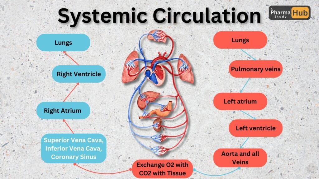
Systemic Circulation: Delivering Oxygen to the Body
Systemic circulation carries oxygen-rich blood from the heart to all parts of the body and brings back deoxygenated blood. Here’s the process:
- The left ventricle pumps oxygen-rich blood into the aorta, the largest artery in the body.
- From the aorta, blood is distributed to arteries, arterioles, and capillaries, reaching every tissue.
- Capillaries facilitate the exchange of oxygen, nutrients, and waste products with the tissues.
- Deoxygenated blood returns to the heart through veins and enters the right atrium via the superior and inferior vena cava.
This circulation ensures that every cell in the body gets the oxygen and nutrients it needs to function while removing waste products.
Coronary Circulation
While the heart pumps blood to the entire body, it also needs its own blood supply to stay strong and function efficiently. Coronary circulation is the system that provides oxygen and nutrients to the heart muscle itself.
Here’s how it works:
- Oxygenated Blood Supply:
- Oxygen-rich blood is delivered to the heart muscle through the coronary arteries, which branch off from the aorta near its base.
- The two main arteries are the left coronary artery (supplying the left side of the heart) and the right coronary artery (supplying the right side).
- Deoxygenated Blood Return:
- After the heart muscle uses the oxygen, deoxygenated blood is collected by the coronary veins.
- These veins drain into the coronary sinus, which empties into the right atrium, allowing the blood to re-enter the pulmonary circulation for oxygenation.
Coronary circulation is the lifeline of the heart itself, ensuring it stays nourished and healthy so it can keep pumping blood for the rest of the body.
Electrical Activity of The Heart
The heart isn’t just a powerful pump; it’s also an incredible electrical system that controls its rhythmic beating. This electrical activity begins at a special group of cells known as the sinoatrial (SA) node, located in the right atrium. Often called the heart’s natural pacemaker, the SA node generates electrical impulses that travel through the heart, coordinating each beat. These signals ensure that the atria and ventricles contract in the right order, maintaining a steady and efficient blood flow throughout the body. This intricate electrical network allows the heart to adapt to different demands, such as during exercise or rest, highlighting the heart’s remarkable ability to keep us alive and active.
Sinoatrial Node(SA)
The sinoatrial node (SA node) is a small group of specialized cells located in the upper wall of the right atrium, near the opening of the superior vena cava. Despite its tiny size, the SA node plays a huge role in keeping the heart beating regularly and rhythmically.
How the SA Node Works:
- Impulse Generation:
- The SA node generates electrical signals, or impulses, at regular intervals.
- These impulses act as the starting point for each heartbeat.
- Setting the Heart Rate:
- The SA node determines the heart rate, adjusting it based on the body’s needs.
- For example, during exercise, the SA node increases the heart rate to pump more blood; during rest, it slows down the rate.
- Spreading the Signal:
- The electrical impulses from the SA node spread through the atria, causing them to contract and push blood into the ventricles.
The SA node is referred to as the natural pacemaker because it sets the rhythm for the entire heart. If it doesn’t function properly, it can lead to irregular heartbeats, requiring an artificial pacemaker to take over the job.
Atrioventricular Node (AV)
The atrioventricular node (AV node) is a small but essential part of the heart’s electrical system, located in the lower part of the right atrium near the interatrial septum. It acts as a critical relay station, ensuring the electrical signals from the sinoatrial (SA) node are transmitted to the ventricles in a controlled and orderly manner.
How the AV Node Works:
- Delays the Signal:
- The AV node introduces a slight delay to the electrical impulses received from the SA node.
- This delay allows the atria to fully contract and push blood into the ventricles before the ventricles contract.
- Relays the Signal:
- After the delay, the AV node sends the impulses down the heart’s electrical pathway (via the Bundle of His and Purkinje fibers) to stimulate the ventricles to contract.
- Backup Pacemaker:
- If the SA node fails to function properly, the AV node can take over as a secondary pacemaker, though it generates signals at a slower rate.
The AV node ensures that the atria and ventricles contract in a coordinated sequence, which is vital for efficient blood flow. Without the AV node’s delay, the heart’s pumping action would become chaotic, reducing its ability to supply blood effectively to the body.
Bundle of HIS and Purkinje Fiber
The Bundle of His and Purkinje fibers are essential components of the heart’s electrical conduction system, responsible for transmitting impulses from the atrioventricular (AV) node to the ventricles. Together, they ensure that the ventricles contract in a synchronized manner, efficiently pumping blood to the lungs and the rest of the body.
Bundle of His: The Signal Carrier
- Location:
- The Bundle of His is located in the upper part of the interventricular septum, the wall between the heart’s two ventricles.
- Function:
- It receives electrical impulses from the AV node and acts as a bridge to pass the signals to the ventricles.
- The bundle splits into two branches:
- Right Bundle Branch: Sends signals to the right ventricle.
- Left Bundle Branch: Sends signals to the left ventricle.
Purkinje Fibers: The Final Pathway
- Location:
- These are thin, specialized fibers located in the walls of the ventricles.
- Function:
- The Purkinje fibers receive impulses from the Bundle of His and distribute them rapidly to the ventricles.
- This causes the ventricles to contract simultaneously, ensuring efficient blood ejection to the lungs and the body.
Fun Facts! About The Heart
15 Fascinating Facts About the Heart
- Heart Size:
- Your heart is about the size of your fist and weighs between 250–350 grams.
- Daily Beast:
- The heart beats approximately 100,000 times a day, pumping blood throughout your body.
- Blood Pumped:
- It pumps around 7,570 liters (2,000 gallons) of blood daily.
- Vascular Length:
- If all your blood vessels were laid end-to-end, they would stretch about 96,000 kilometers—enough to circle the Earth more than twice!
- Fastest Muscle:
- The heart is the hardest-working muscle, starting to pump blood just weeks after conception and continuing for a lifetime.
- Dual Pump System:
- The heart acts as two pumps: the right side sends blood to the lungs, and the left side sends blood to the rest of the body.
- Heartbeat Control:
- The heart generates its own electrical impulses and can continue beating even when removed from the body, as long as it has an oxygen supply.
- Fastest Heartbeat:
- A newborn’s heart beats much faster than an adult’s, averaging 120–160 beats per minute compared to 60–100 bpm in adults.
- Lifespan Beats:
- Over a lifetime, the heart beats about 3 billion times on average.
- Unique Rhythm:
- Each person’s heartbeat rhythm is unique, much like fingerprints.
- Sound of Heartbeat:
- The “lub-dub” sound is caused by the closing of heart valves during the cardiac cycle.
- Heart Blood Supply:
- The heart muscle itself is nourished by its own blood supply through the coronary arteries.
- Exercise Benefits:
- Regular exercise strengthens the heart, allowing it to pump more efficiently and lower resting heart rate.
- Emotional Influence:
- Strong emotions like love, fear, or excitement can temporarily alter your heart rate due to the brain-heart connection.
- Heart in Numbers:
- The average adult heart pumps about 70 milliliters of blood per beat, moving 5 liters of blood every minute!
The heart is truly an amazing organ, tirelessly working to keep us alive and functioning every second of the day!
Conclusion
The heart is more than just a muscle; it’s the engine that powers life. From its intricate anatomy to its efficient physiology, every beat is a testament to its remarkable design. Whether it’s pumping oxygen-rich blood to every corner of the body or adapting to our ever-changing needs, the heart works tirelessly to keep us alive.
Understanding how the heart functions not only deepens our appreciation for this vital organ but also highlights the importance of keeping it healthy through regular exercise, a balanced diet, and a stress-free lifestyle.
As we explore the wonders of the cardiovascular system, let’s remember that taking care of our heart isn’t just about extending life—it’s about living it to the fullest. Stay tuned for more insights into the amazing world of the human body!
FAQs
The heart’s main function is to pump blood throughout the body, delivering oxygen and nutrients to cells while removing waste products like carbon dioxide.
The heart has four chambers: two atria (upper chambers) and two ventricles (lower chambers).
Arteries carry oxygen-rich blood away from the heart to the body, while veins bring oxygen-poor blood back to the heart.
The “lub-dub” sound is caused by the closing of heart valves during the cardiac cycle. The “lub” is from the atrioventricular valves closing, and the “dub” is from the semilunar valves closing.
The SA node is the heart’s natural pacemaker. It generates electrical signals that control the heart’s rhythm and regulate the heartbeat.
Coronary circulation provides blood supply to the heart muscle itself through the coronary arteries and veins, ensuring the heart gets the oxygen and nutrients it needs to function.
Exercise strengthens the heart, improves its efficiency, lowers resting heart rate, and promotes better blood circulation.
Blood pressure is the force of blood pushing against artery walls as the heart pumps. Maintaining healthy blood pressure is crucial to prevent strain on the heart and reduce the risk of heart disease.
A heart attack occurs when blood flow to part of the heart muscle is blocked, usually by a clot in the coronary arteries. This can damage or destroy part of the heart muscle.
To keep your heart healthy, eat a balanced diet, exercise regularly, avoid smoking, manage stress, and maintain healthy blood pressure and cholesterol levels.